1. Cobalt Sulfate, Heptahydrate
2. Cobaltous Sulfate
1. Cobaltous Sulfate
2. Cobalt(2+) Sulfate
3. 10124-43-3
4. Cobalt(ii) Sulfate
5. Cobalt Sulphate
6. Cobalt Brown
7. Cobalt Monosulfate
8. Cobalt(ii) Sulphate
9. Cobalt (2+) Sulfate
10. Cobalt Sulfate (1:1)
11. Cobalt(2+);sulfate
12. Cobalt(ii) Sulfate (1:1)
13. Sulfuric Acid, Cobalt(2+) Salt (1:1)
14. Cobalt(+2) Cation Sulfate
15. H7965x29hx
16. 139939-65-4
17. Cobalt Sulfate (coso4)
18. Hsdb 240
19. Cobaltous Sulfate Salt (1:1)
20. Einecs 233-334-2
21. Unii-h7965x29hx
22. Coso4
23. Cobaltous Sulphate
24. Ec 233-334-2
25. Cobalt(2+) Sulphate
26. Cobalt( Cento) Sulfate Hydrate
27. Cobalt(ii) Sulfate, Anhydrous
28. Cobaltous Sulfate [mi]
29. Cobalt Sulfate [who-dd]
30. Cobaltous Sulfate [hsdb]
31. Dtxsid1031042
32. Chebi:53470
33. Cobalt Sulphate (1:1)
34. Mfcd00010943
35. Akos015903535
36. C17383
37. A800357
38. Q411214
| Molecular Weight | 155.00 g/mol |
|---|---|
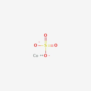
| Molecular Formula | CoO4S |
| Hydrogen Bond Donor Count | 0 |
| Hydrogen Bond Acceptor Count | 4 |
| Rotatable Bond Count | 0 |
| Exact Mass | 154.884923 g/mol |
| Monoisotopic Mass | 154.884923 g/mol |
| Topological Polar Surface Area | 88.6 Ų |
| Heavy Atom Count | 6 |
| Formal Charge | 0 |
| Complexity | 62.2 |
| Isotope Atom Count | 0 |
| Defined Atom Stereocenter Count | 0 |
| Undefined Atom Stereocenter Count | 0 |
| Defined Bond Stereocenter Count | 0 |
| Undefined Bond Stereocenter Count | 0 |
| Covalently Bonded Unit Count | 2 |
The effects of cobalt sulfate administered to pregnant C57BI mice, OFA-SD rats, and New Zealand rabbits was studied on fetal and postnatal offspring. Cobalt concentration in the maternal blood was increased in proportion to the administered doses. Cobalt crossed the placenta and appeared in the fetal blood and amniotic fluid. Regardless of the administered dose of cobalt sulfate, cobalt concentration in the blood peaked 2 hr after administration.
PMID:11261899 Szakmary E et al; J Toxicol Environ Health A 62 (5): 367-86 (2001)
Sheep given a single dose of hydrated cobalt sulfate were killed. ...The livers contained 400-1200 ppm of cobalt. In cattle dying after a massive overdose of hydrated cobalt sulfate livers contained 5-300 ppm, kidneys 30-200 ppm. /Hydrated cobalt sulfate/
Clarke, M. L., D. G. Harvey and D. J. Humphreys. Veterinary Toxicology. 2nd ed. London: Bailliere Tindall, 1981., p. 44
Following longer-term exposure (8 weeks) to cobalt sulfate in the diet, exposed rats showed a 30-fold increase in the cobalt concentration in the myocardium, a 26-fold increase in the concentration in the soleus muscle, and a 100-fold increase in the concentration in serum compared with nonexposed controls. Long-term oral exposure of rats to cobalt chloride resulted in significantly increased levels of cobalt in the liver, kidney, muscle, brain, and testes of treated rats.
DHHS/ATSDR; Toxicological Profile for Cobalt p.130 (PB/93/110724/AS) (April 2004)
Cobalt metabolism and toxicology are summarized. The biological functions of cobalt are updated in the light of recent understanding of cobalt interference with the sensing in almost all animal cells of oxygen deficiency (hypoxia). Cobalt (Co(2+)) stabilizes the transcriptional activator hypoxia-inducible factor (HIF) and thus mimics hypoxia and stimulates erythropoietin (Epo) production, but probably also by the same mechanism induces a coordinated up-regulation of a number of adaptive responses to hypoxia, many with potential carcinogenic effects. This means on the other hand that cobalt (Co(2+)) also may have beneficial effects under conditions of tissue hypoxia, and possibly can represent an alternative to hypoxic preconditioning. Cobalt is acutely toxic in larger doses, and in mammalian in vitro test systems cobalt ions and cobalt metal are cytotoxic and induce apoptosis and at higher concentrations necrosis with inflammatory response. Cobalt metal and salts are also genotoxic, mainly caused by oxidative DNA damage by reactive oxygen species, perhaps combined with inhibition of DNA repair. Of note, the evidence for carcinogenicity of cobalt metal and cobalt sulfate is considered sufficient in experimental animals, but is as yet considered inadequate in humans. Interestingly, some of the toxic effects of cobalt (Co(2+)) have recently been proposed to be due to putative inhibition of Ca(2+) entry and Ca(2+)-signaling and competition with Ca(2+) for intracellular Ca(2+)-binding proteins. The tissue partitioning of cobalt (Co(2+)) and its time-dependence after administration of a single dose have been studied in man, but mainly in laboratory animals. Cobalt is accumulated primarily in liver, kidney, pancreas, and heart, with the relative content in skeleton and skeletal muscle increasing with time after cobalt administration. In man the renal excretion is initially rapid but decreasing over the first days, followed by a second, slow phase lasting several weeks, and with a significant long-term retention in tissues for several years. In serum cobalt (Co(2+)) binds to albumin, and the concentration of free, ionized Co(2+) is estimated at 5-12% of the total cobalt concentration. In human red cells the membrane transport pathway for cobalt (Co(2+)) uptake appears to be shared with calcium (Ca(2+)), but with the uptake being essentially irreversible as cobalt is effectively bound in the cytosol and is not itself extruded by the Ca-pump. It is tempting to speculate that this could perhaps also be the case in other animal cells. If this were actually the case, the tissue partitioning and biokinetics of cobalt in cells and tissues would be closely related to the uptake of calcium, with cobalt partitioning primarily into tissues with a high calcium turn-over, and with cobalt accumulation and retention in tissues with a slow turn-over of the cells. The occupational cobalt exposure, e.g. in cobalt processing plants and hard-metal industry is well known and has probably been somewhat reduced in more recent years due to improved work place hygiene. Of note, however, adverse reactions to heart and lung have recently been demonstrated following cobalt exposure near or slightly under the current occupational exposure limit. Over the last decades the use of cobalt-chromium hard-metal alloys in orthopedic joint replacements, in particular in metal-on-metal bearings in hip joint arthroplasty, has created an entirely new source of internal cobalt exposure. Corrosion and wear produce soluble metal ions and metal debris in the form of huge numbers of wear particles in nanometric size, with systemic dissemination through lymph and systemic vascular system. This may cause adverse local reactions in peri-prosthetic soft-tissues, and in addition systemic toxicity. Of note, the metal nanoparticles have been demonstrated to be clearly more toxic than larger, micrometer-sized particles, and this has made the concept of nanotoxicology a crucial, new discipline. As another new potential source of cobalt exposure, suspicion has been raised that cobalt salts may be misused by athletes as an attractive alternative to Epo doping for enhancing aerobic performance. The cobalt toxicity in vitro seems to reside mainly with ionized cobalt. It is tempting to speculate that ionized cobalt is also the primary toxic form for systemic toxicity in vivo. Under this assumption, the relevant parameter for risk assessment would be the time-averaged value for systemic cobalt ion exposure that from a theoretical point of view might be obtained by measuring the cobalt content in red cells, since their cobalt uptake reflects uptake only of free ionized cobalt (Co(2+)), and since the uptake during their 120 days life span is practically irreversible. This clearly calls for future clinical studies in exposed individuals with a systematic comparison of concurrent measurements of cobalt concentration in red cells and in serum.
PMID:22732165 Simonsen LO et al; Sci Total Environ 432: 210-5 (2012)
Cellular metabolism depends on the availability of oxygen and the major regulator of oxygen homeostasis is hypoxia-inducible factor 1 (HIF-1), a highly conserved transcription factor that plays an essential role in cellular and systemic homeostatic responses to hypoxia. HIF-1 is a heterodimeric transcription factor composed of hypoxia-inducible HIF-1alpha and constitutively expressed HIF-1beta. Under hypoxic conditions, the two subunits dimerize, allowing translocation of the HIF-1 complex to the nucleus where it binds to hypoxia-response elements (HREs) and activates expression of target genes implicated in angiogenesis, cell growth, and survival. The HIF-1 pathway is essential to normal growth and development, and is involved in the pathophysiology of cancer, inflammation, and ischemia. Thus, there is considerable interest in identifying compounds that modulate the HIF-1 signaling pathway. To assess the ability of environmental chemicals to stimulate the HIF-1 signaling pathway, we screened a National Toxicology Program collection of 1408 compounds using a cell-based beta-lactamase HRE reporter gene assay in a quantitative high-throughput screening (qHTS) format. Twelve active compounds were identified. These compounds were tested in a confirmatory assay for induction of vascular endothelial growth factor, a known hypoxia target gene, and confirmed compounds were further tested for their ability to mimic the effect of a reduced-oxygen environment on hypoxia-regulated promoter activity. Based on this testing strategy, three compounds (o-phenanthroline, iodochlorohydroxyquinoline, cobalt sulfate heptahydrate) were confirmed as hypoxia mimetics, whereas two compounds (7-diethylamino-4-methylcoumarin and 7,12-dimethylbenz(a)anthracence) were found to interact with HIF-1 in a manner different from hypoxia. /Cobalt sulfate heptahydrate/
PMID:19502547 Full text: https://www.ncbi.nlm.nih.gov/pmc/articles/PMC2910898 Xia M et al; Toxicol Sci 112 (1): 153-6 (2009)